tree in bud opacities
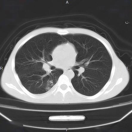
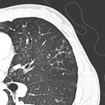
1 5 6 7 8 9 10 11 12. 8081 On CT the tree-in-bud pattern manifests as small 24 mm centrilobular well-defined nodules connected to linear.
2
Nuclear Medicine 53 years experience.
. The list of the most frequent differential diagnoses for tree-in-bud sign includes infections with Mycobacterium tuberculosis nontuberculous mycobacteria and other bacterial fungal or viral pathogens. 87 rows Uncommonly this pattern can be seen in other entities that cause luminal impaction bronchiolar dilatation or wall thickening including cystic fibrosis immune deficiency inflammatory bowel disease and diffuse panbronchiolitis. Ad Get the Latest On The First Signs of Lung Cancer In This Article.
Originally and still often thought to be specific to endobronchial Tb the sign is actually non-specific and is the manifestation of pus mucus fluid or other. Tree in bud opacification refers to a sign on chest CT where small centrilobular nodules and corresponding small branches simulate the appearance of the end of a branch belonging to a tree that is in bud. TIB opacities typically show branching configurations from secondary pulmonary lobules with sparing of subpleural.
The purpose of this study was to determine the relative frequency of causes of TIB opacities and identify patterns of disease associated with TIB opacities. Other causes could be immunological congenital and idiopathic disorders as well as aspiration or inhalation of. Sarcoidosis another common disease typically shows small nodules in perilymphatic distribution.
Multiple causes for tree-in-bud TIB opacities an imaging pattern usually seen on chest CT have been reported. However in some cases nodules occurring in relation to centrilobular arteries may mimic the appearance of the tree-in-bud pattern. Originally and still often thought to be specific to endobronchial Tb the sign is actually non-specific and is the manifestation of pus mucus fluid or other.
However BAC can occasionally show tree-in-bud pattern ground-glass opacities or crazy-paving pattern. Respiratory infections 72 with TB. Created for people with ongoing healthcare needs but benefits everyone.
1 The tree-in-bud sign is a nonspecific imaging finding that implies impaction within bronchioles the smallest airway passages in the lung. Multiple causes for tree-in-bud TIB opacities have been reported. 1 2 3 4 Reported causes include infections aspiration and a variety of inflammatory conditions.
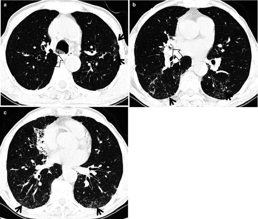
Tree-in-bud opacities with false-positive Gaffky score and diffuse aspiration bronchiolitis. However after listening to patients voice and reviewing the images on CT thorax the diagnosis was confirmed as aspiration bronchiolitis. INTRODUCTION Tree-in-bud TIB opacities are a subset of centrilobular nodules.
The differential diagnosis of tree-in-bud nodules includes infection and aspiration the two most common causes as well as congenital airway diseases allergic bronchopulmonary aspergillosis follicular bronchiolitis panbronchiolitis intravenous injection of foreign bodies and other causes. TIB opacities typically show branching configurations from secondary pulmonary lobules with sparing of subpleural lungs on CT thorax. These are due to filling of the distal bronchioles and involvement of the adjacent alveoli most often caused by infectious bronchiolitis bronchitis and aspiration.
Tree-in-bud TIB opacities are a subset of centrilobular nodules. While the tree-in-bud appearance usually represents endobronchial spread of infection given the closeness of small pulmonary arteries and small airways sharing branching morphology-bronchovascular bundle a rarer cause of the tree-in-bud sign is infiltration of the small pulmonary arteries or axial interstitium 367. Provisional diagnosis of pulmonary tuberculosis was made and was referred to the respiratory team.
The differential for this finding includes malignant and inflammatory etiologies either infectious or sterile. The tree-in-bud sign is a common finding in HRCT scans. Tree-in-bud TIB opacities are a common imaging finding on thoracic CT scan.
Contrast-enhanced CT computed tomography thorax revealed tree-in-bud TIB opacities. However to our knowledge the relative frequencies of the causes have not been evaluated. These small clustered branching and nodular opacities represent terminal airway mucous impaction with adjacent peribronchiolar inflammation.
Tree-in-bud opacities appear as tiny centrilobular branching structures on CT most often in the lung periphery which resemble budding trees Figure 18-4. Tree-in-bud opacities detected after aspiration should be considered DAB rather than mycobacterial infection. Tree in bud opacification refers to a sign on chest CT where small centrilobular nodules and corresponding small branches simulate the appearance of the end of a branch belonging to a tree that is in bud.
In radiology the tree-in-bud sign is a finding on a CT scan that indicates some degree of airway obstruction. CT finding of centrilobular nodules with TIB opacities was first described in pulmonary tuberculosis and is considered highly predictive of.
2

Tree In Bud Pattern Radiology Case Radiopaedia Org
Tree In Bud Sign Lung Radiology Reference Article Radiopaedia Org
2

Tree In Bud Sign Lung Radiology Reference Article Radiopaedia Org

Pdf Tree In Bud
View Of Tree In Bud The Southwest Respiratory And Critical Care Chronicles

Tree In Bud Pattern Pulmonary Tb Eurorad

Tree In Bud Sign And Bronchiectasis Radiology Case Radiopaedia Org

Chest Ct With Multifocal Tree In Bud Opacities Diffuse Bronchiectasis Download Scientific Diagram

Pdf Tree In Bud Semantic Scholar
Tree In Bud Sign Lung Radiology Reference Article Radiopaedia Org

Hrct Scan Of The Chest Showing Diffuse Micronodules And Tree In Bud Download Scientific Diagram
2

Tree In Bud Sign Lung Radiology Reference Article Radiopaedia Org
Tree In Bud Sign Radiology Key

Tree In Bud Pattern Radiology Case Radiopaedia Org
2
2